Our range of imaging services includes:
- Workers Compensation related imaging
- CT Scan
- Angiography
- Angiogram Vessel analysis
- Cardiac Imaging (CTCA)
- X-Ray
- Ultrasound
- Obstetric Ultrasound
- Elastography – liver, breast, and soft tissue
- 3D Mammography
- Injections/Procedures/Biopsy
- Image Guided Biopsy
- Fizz Study (Stomach)
- Virtual CT Colonoscopy (Fly-through)
- Discogram
- Arthrogram
- MRI
- 4D Bub-Lab
Book now for fast turnaround services and bulk-billing on all Medicare eligible examinations.
Workers Compensation related imaging
Our medical imaging specialists have extensive experience in workers’ compensation injuries, providing services to both corporate and insured clients. Working in partnership with allied workers’ compensation specialists and clinics across South West and Western Sydney, we are able to streamline and accelerate the management process.
Westside Medical Imaging will work with you to:
- Organise pre-approvals (patients will need to have a claim number)
- Book any appointments, including Interventional Procedures
- Provide fast, timely appointments where quick turnaround is required
- Deliver professional reports by our Specialist Radiologist.
We offer a complete pathway, from clinical and radiological diagnosis to the safe treatment of injuries or conditions using image-guided therapeutic injections. With some of the most advanced medical imaging equipment available, we deliver accurate diagnoses promptly and efficiently, helping patients return to work sooner.
We also support the Jobfit Program and Pre-Employment Scans.
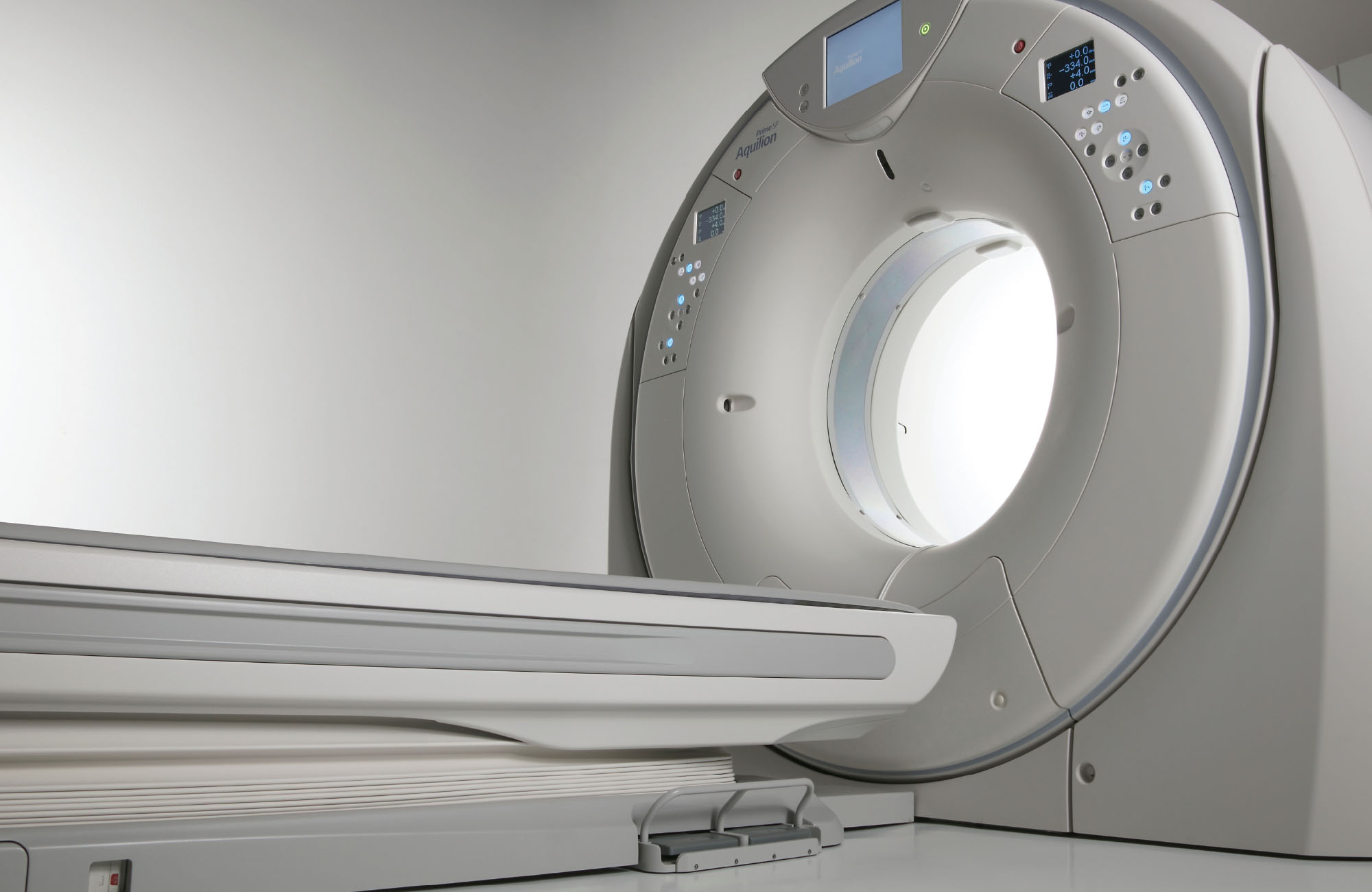
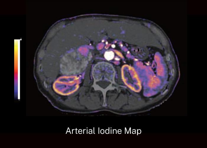
CT Scan
Our CT scanner features the industry’s thinnest detector and collimator elements (0.5mm) and widest bore size (78cm) in Australia, to deliver high resolution images at genuine ultra low-doses (up to 82% reduction of radiation exposure).
CT Iodine Mapping (spectral imaging) increases the diagnostic information available for any routine multiphase body protocol by extracting the iodine density on each phase, without increasing radiation exposure.
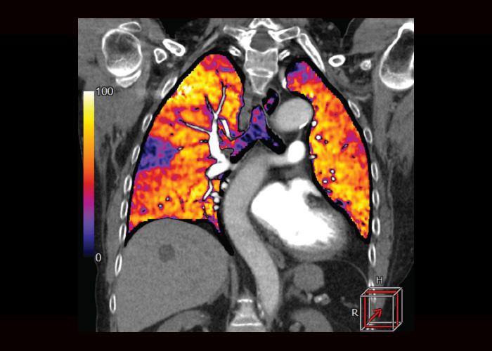
Angiography
Angiography is used to check the health of your blood vessels and how blood flows through them. It can help to diagnose or investigate several problems affecting blood vessels. If detected, this could mean you’re at risk of having a stroke or heart attack, pulmonary emboli, renal infarction, or a diabetic foot.
Our team is highly trained and experienced in performing all types of angiography, ensuring accurate diagnosis and the highest standards of patient care.
Angiogram vessel analysis
CT imaging that provides a 3D rotatable reconstruction of the vessels from all dimensions in order to detect any stenosis of the arteries and additional pathologies such as dissection, aneurysm, TIA or stoke.
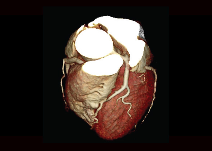

Cardiac Imaging (CTCA)
At Westside Medical Imaging, we provide a variety of advanced cardiac CT imaging, including CT coronary angiography (CTCA), myocardial perfusion imaging, functional analysis, calcium score of coronary arteries or valves, aortography, and dual or triple vessel rule out scans to precisely assess vessel patency, functions and relevant structural abnormalities of the heart vessels, valves and walls.
Our premium-grade CT scanners are equipped with real time or post processing AI reconstruction for dose- and noise reductions, ensuring the highest image quality with the lowest possible radiation exposure. We work in close collaboration with experienced cardiologists to deliver accurate diagnoses and a tailored care to every patient, helping your doctor detect and manage heart disease early for the best possible outcomes.
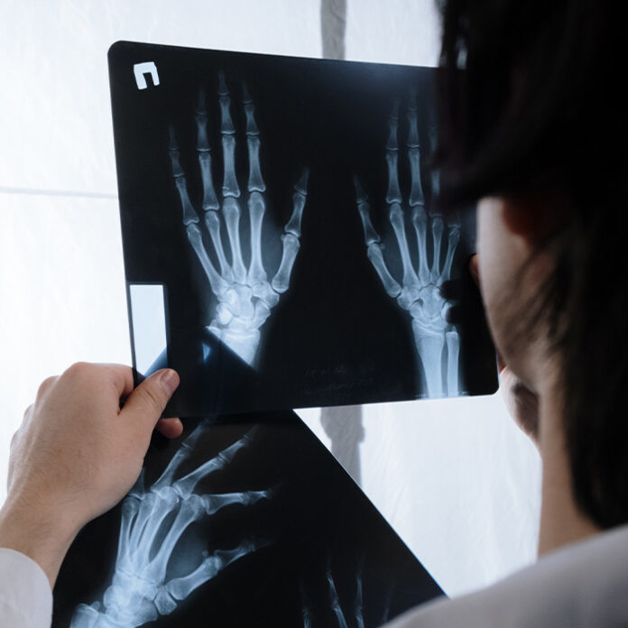
X-Ray
The latest Shimadzu digital x-ray system provides our local community with high resolution imaging at a reduced radiation dose, enabling quick diagnoses of broken bones and joint pathology.
No bookings are required for X-Rays, walk-in appointments are welcomed.
Ultrasound
An ultrasound scan, also called sonography or diagnostic medical sonography, is an imaging method using high-frequency sound waves to produce images of structures within the body. The images can provide valuable information for diagnosing and treating a variety of diseases and conditions, yet is most commonly used on a pregnant woman to examine the fetus.
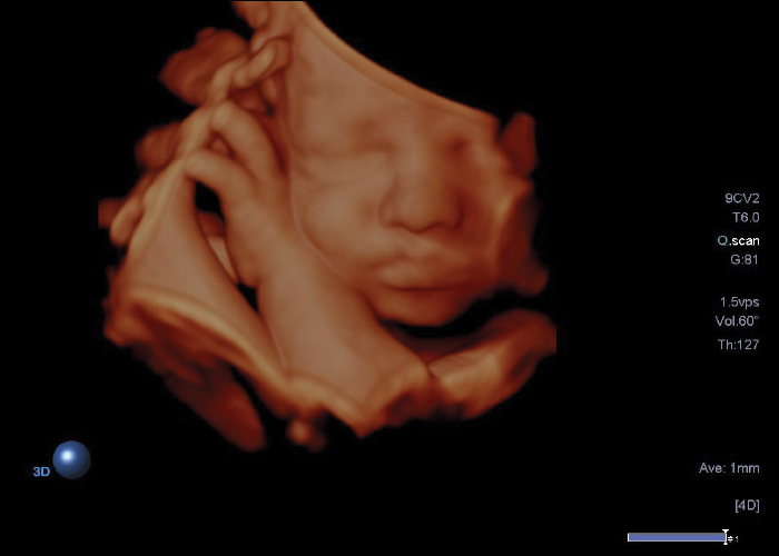
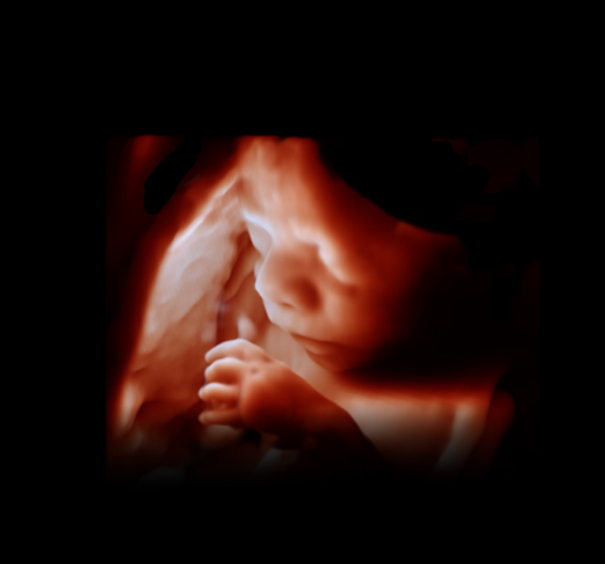
Obstetric Ultrasound
We offer advanced 3D and 4D obstetric ultrasound imaging, performed using the advanced Philips imaging technology for exceptional clarity and fine detail. Our highly experienced senior male and female sonographers provide accurate, reliable results and compassionate care. We are committed to delivering high-quality, personalised care throughout every stage of your pregnancy.
Elastography – liver, breast, and soft tissue
Shear-wave elastography (SWE) is a non-invasive assessment of tissue fibrosis, chronic liver disease (including NAFLD), and neoplastic lesions in the liver, breast and other soft tissues. Radiation or damage/ pain free acoustic impulse offers a substantial disease-specific information of tissue stiffness, segregates the extent of fibrosis and assist in characterizing tumors (grade, extension and mobility).
3D Mammography
3D mammography (Breast tomosynthesis) is performed with a single short breath hold and an X-ray tube pivoting in an arc between 15-60 degrees. It is reported that 3D mammography increases breast cancer detection rate, ranging from 1.2 to 4.6 per 1000 examination (approximate increase of 40%) compared to conventional 2D mammography and improves specificity across all breast densities.
Interventional Procedures
We offer a comprehensive range of image-guided procedures, including steroid, hyaluronic acid (Euflexxa), and Platelet rich plasma (PRP) injections, as well as biopsy and aspiration services. Our focus is on delivering minimally invasive treatments, performed by highly experienced radiologists dedicated to achieving the best possible patient outcomes.
Our lead radiologist, Dr Tom is recognised for his precision, advanced technical expertise, and commitment to patient comfort. With experience in over 15,000 successful procedures, he ensures each patient receives compassionate care with minimal pain and the best possible treatment results. Refer to our Google reviews for his professional procedure details.
Consultation sessions are available for individualised assessment and tailored treatment planning (referral required).
Platelet-Rich Plasma (PRP)
Injections uses a concentration of your own blood cells and plasma, which reduces the risk of an allergic reaction or adverse event, into an injured area, to optimize the initial inflammatory response and accelerate the healing of damaged tendons, ligaments, muscles and joints.
A key advantage of PRP injections is to reduce recovery time and prevent a recurrence after non-surgical injuries, those not limited to but including tennis elbow, rotator cuff tear, or Achilles tendonitis.
After the procedure, you may experience increasing soreness and bruising at the injection site to some extent and should limit the use of the treated area for the next couple of days to allow the area to absorb all the platelets properly.
The benefits of PRP injections will be maximized between four to six weeks to be noticeable but will continue to aid in further healing for anywhere up to six to nine months.
Image Guided Biopsy
An image-guided biopsy is a procedure done under ‘image guidance’ which removes a small sample of tissue or cells for examination under a microscope to see if cancer or other abnormalities are present. The type of imaging used for your biopsy will depend on the location of the abnormality and how it is best seen.
Fizz Study
Specialised test that provides 3D imaging of the stomach size and orientation to assist planning for potential revisional surgeries which include gastric sleeves, bands and bypass for weight loss.
Virtual Colonoscopy (EC function)
Our advanced technology virtually clears bowel residues for optimal viewing, helping to detect polyps early and support the early detection of colon cancer. The procedure is non-invasive, and you can rest assured that our experienced team will take care of you, ensuring a comfortable and reassuring experience.

Discogram
A discogram is a type of CT guided diagnostic interventional procedure which is performed for the assessment of the severity of spinal discomfort which may be due to abnormalities with the intervertebral disc/s. The procedure aids with identifying whether your back pain is due to disc disruption and if this is the case, it allows your doctor to identify which disc level/s are involved and what treatment may be beneficial for your pain management.
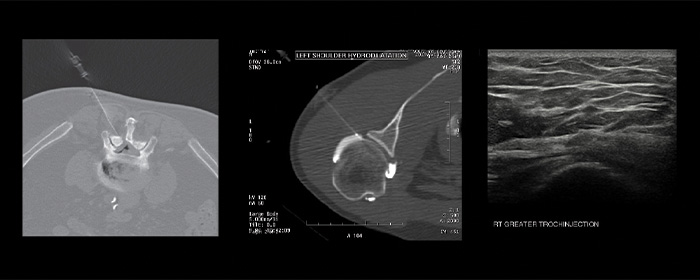
Arthrogram
An arthrogram is a medical imaging procedure which aids with the diagnosis of subtle abnormalities in your joints. The most commonly targeted joints for an arthrogram include the shoulder, knee, hip, elbow and hand/wrist. A CT scanner is used to target the location for which the arthrogram will be performed. This procedure is commonly requested by your doctor prior to an MRI scan for them to visualise how contrast spreads around the joint space, which assists with the diagnosis and treatment of certain joint conditions which may be causing your ongoing pain/discomfort. Often, an arthrogram is requested if other forms of medical imaging (e.g., X-ray, Ultrasound, MRI, CT) do not provide sufficient information to allow for an adequate diagnosis, treatment or management plan to be made.
MRI
Westside Medical Imaging will be providing the first Zero helium MRI scanner in Wetherill Park and surrounding suburbs soon. We will offer variety of conventional and advanced MR imaging including Neuro, Angiography, Head & Neck, Spinal, Cardiac, Abdomen/pelvis, MSK, and Prostate examinations using the latest superconductive MR scanner.
The scanner will be equipped with a patient-friendly design which provides comfort and versatility by offering the wide bore and a special headset for claustrophobic patients, and ultra-fast scans with AI technology.
Book now for fast turnaround services and bulk-billing on all Medicare eligible examinations.
Wetherill Park

Bonnyrigg

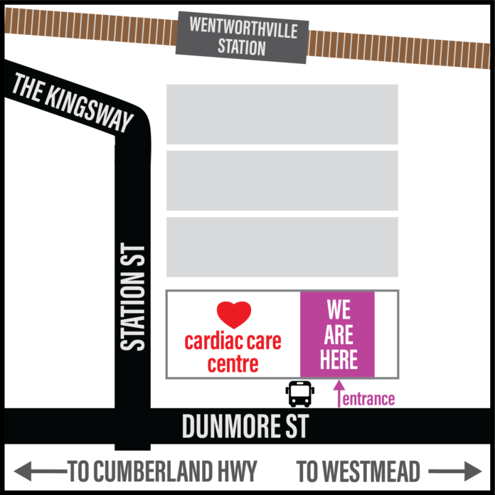
Wentworthville